Extraordinary Animals in the Womb is a Stunning Collection
References: channel.nationalgeographic & thisblogrules
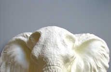
These mind-blowing images of tiny animal fetuses were made for the National Geographic documentary ‘Extraordinary Animals in the Womb’ and are the product of three-dimensional ultrasound scanning, micro camera photography and computer-generated modeling technology.
To capture these phenomenal images, the filmmakers of ‘Extraordinary Animals in the Womb’ scanned pregnant animals’ wombs and had a model-making team painstakingly recreate every detail. These photos are an unparalleled window into the early life stages of sharks, cats, dogs, elephants and even penguins in vivid detail.
To capture these phenomenal images, the filmmakers of ‘Extraordinary Animals in the Womb’ scanned pregnant animals’ wombs and had a model-making team painstakingly recreate every detail. These photos are an unparalleled window into the early life stages of sharks, cats, dogs, elephants and even penguins in vivid detail.
Trend Themes
1. 3D Ultrasound Scanning - Using 3D ultrasound scanning technology to capture detailed images of animal embryos could revolutionize the field of embryology and provide valuable insights into prenatal development.
2. Micro Camera Photography - The use of micro camera photography in capturing images of animal fetuses in the womb presents new opportunities for studying and understanding the intricacies of animal development.
3. Computer-generated Modeling - The application of computer-generated modeling technology in recreating animal fetuses allows for a level of detail and accuracy that was previously impossible, opening up possibilities for further scientific research and educational purposes.
Industry Implications
1. Embryology - The field of embryology could benefit from the use of 3D ultrasound scanning, micro camera photography, and computer-generated modeling to advance the understanding of prenatal development.
2. Documentary Filmmaking - The use of advanced imaging technologies such as 3D ultrasound scanning, micro camera photography, and computer-generated modeling in documentary filmmaking can provide stunning visuals and educational content.
3. Education - The combination of 3D ultrasound scanning, micro camera photography, and computer-generated modeling can be leveraged in educational settings to enhance the study of biology and animal development.
6.6
Score
Popularity
Activity
Freshness

